What is short sight (myopia)?
Short sight occurs when light coming from distant objects is ‘overfocused’, so that the point of focus is in front of the retina. It occurs because either the eyeball is too long, or because the cornea is too curved. Despite maximum flattening of the lens, the eye is not able to focus the light rays further back and on to the retina.
Light coming from near objects requires a stronger focusing activity anyway, so in myopia light from near objects is more likely to be focused in the right place.
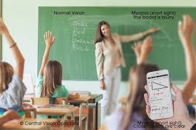
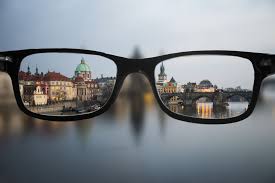
People with short sight are not able to see distant objects clearly. Short sight or near sight mean exactly what the terms suggest. You are sighted (you can see), near (short) distance objects. Near objects (for example, when reading a book) can often be seen well. This is because when looking at near objects, the light rays come into the eye going slightly outwards. These will focus further back in the eye than light rays that come in straight from distant objects.
The diagram above shows the differences in focusing between a normal and a short-sighted (myopic) eye.
What causes short sight (myopia)?
Short sight tends to happen in children and young teenagers. It often runs in families. Temporary short-sightedness can also occur with certain illnesses – for example, in diabetes.
What are the symptoms of short sight (myopia)?
The main symptom is a difficulty with distance vision. The earlier short sight starts, the more severe it is likely to become. By the time early adulthood is reached, the level of short sight has usually reached its peak. This means that the vision does not generally become any worse.
Some children do not realise that their vision is not as good as it should be. They may be able to read books and do close work well. However, seeing distant objects such as the board at school may become difficult. They may think this is normal and not tell anyone. Schoolwork may suffer for a while before the condition is identified and treatment provided.
Children usually have a routine preschool or school-entry vision check. Your child’s teacher may notice that children are having difficulties in class reading the board. If you suspect your child has problems with his or her sight, you should arrange for an eyesight test with an optician who is happy to assess children. For young children and toddlers, your GP may be able to make arrangements for a sight test. Sight tests are free for children.
What is anisometropia?
Very few people are born with two eyes of identical optical power, but the brain manages to compensate and it’s usually unnoticeable.
However, when a person has anisometropia, the difference in vision between their two eyes is significant and will interfere with normal binocular vision. In practice, they will see a smaller image in one eye and a larger image in the other eye. The result is that their overall vision is often blurred.
Another potential outcome from anisometropia is amblyopia (lazy eye), which can occur if one eye has blurred vision for some time and becomes permanently weaker.
Sometimes anisometropia can be present at birth, although frequently it won’t become apparent until later in life. It has been estimated that around six percent of all children aged between six and eighteen suffer from this visual condition.
Types of anisometropia
There are three types of anisometropia:
- Simple anisometropia. This is when one eye is affected while the other eye has no refractive error (or spectacle prescription). The affected eye can either be hyperopic (long-sighted) or myopic (short-sighted).
- Compound anisometropia. This is when both eyes are myopic (short-sighted), although there will be a significant difference in their refractive errors (or spectacle prescriptions). This causes one eye to see a more blurred image than the other.
- Mixed anisometropia. This is when both eyes have appreciable refractive errors, with one eye myopic and the other hyperopic.
Symptoms of anisometropia
There are a number of potential symptoms, including:
- Amblyopia (lazy eye). Usually, this is when reduced refractive power in one eye causes a lack of visual stimulation that results in insufficient information being transmitted through the optic nerve to the brain
- Strabismus (crossed eyes). When a patient is unable to align both eyes. This lack of coordination prevents both eyes being able to focus on the same point in space
- Diplopia (often known as double vision). The result includes:
- Eyestrain
- Headaches
- Nausea
- Light sensitivity
- Tiredness
- Dizziness.
Causes
Even people who have normal vision can have up to 5% difference in the refractive power of each eye. However, those with a 5–20% difference will experience uneven vision (anisometropia). Causes include defects in the eye at childbirth as well as uneven size of the two eyes.

Visual acuity, eyesight and vision: What’s the difference?
Visual acuity
Visual acuity is the clarity of your eyesight, measured by your ability to identify letters or numbers on a standardized eye-chart from a specific viewing distance.
Visual acuity is a static measurement, meaning you are sitting still during the testing and the letters or numbers you are viewing also are stationary.
Visual acuity also is tested under high contrast conditions — typically, the letters or numbers on the eye chart are black, and the background of the chart is white.
Although visual acuity testing is very useful to determine the relative clarity of your eyesight in standardized conditions, it isn’t predictive of the quality of your vision in all situations.
For example, it can’t predict how well you would see:
- Objects that are similar in brightness to their background
- Colored objects
- Moving objects
Three major physical and neurological factors determine visual acuity:
- How accurately the cornea and lens of the eye focus light onto the retina
- The sensitivity of the nerves in the retina and vision centers in the brain
- The ability of the brain to interpret information received from the eyes
Only light that is focused on a very small and highly sensitive portion of the central retina (called the macula) influences visual acuity measurements obtained during an eye exam.
Eyesight
The exact definition of “eyesight” is difficult to pin down. Depending on which dictionary or other resource you check, it can mean “ability to see,” “the sense of seeing,” “vision,” “range of sight” or “view.” Often, the terms “eyesight” and “visual acuity” are used interchangeably.
Vision
Vision is a broader term than visual acuity or eyesight. In addition to clarity of sight or simply a description of the ability to see, the term “vision” all interactions between the eyes and the brain, and all neurological processes that take place in the brain to make the sense of vision possible.
Also, unlike simple eyesight or Snellen (high contrast) visual acuity, measures of vision include contrast sensitivity, the ability to track moving objects with smooth and accurate eye movements, color vision, depth perception, focusing speed and accuracy, and more.
Because of the broader nature of the word “vision,” what is commonly called “20/20 vision” should really be called “20/20 visual acuity” or “20/20 eyesight.”
What is hypermetropia?
Hypermetropia, sometimes called hyperopia, is the term used to define being longsighted.
If you are hypermetropic, the image of a nearby object is formed behind the retina. This means that light is focused too far back in the eye, causing things which are close up to appear blurred.
How is it treated?
Both myopia and hypermetropia can be easily corrected by the Optometrist at your local Vision Express, using prescription glasses or contact lenses specifically designed to counteract their effects.
Shortsightedness is corrected using a concave (curved inwards) lens which is placed in front of a myopic eye, moving the image back to the retina and making it clearer.
Longsightedness is corrected using a convex (outward facing) lens. This is placed in front of a hypermetropic eye, moving the image forward and focusing it correctly on the retina.
Astigmatism
Astigmatism is a common vision condition that causes blurred vision. It occurs when the cornea (the clear front cover of the eye) is irregularly shaped or sometimes because of the curvature of the lens inside the eye.
Astigmatism is an irregularly shaped cornea or lens that prevents light from focusing properly on the retina, the light-sensitive surface at the back of the eye. The surface of the cornea is shaped more like a football instead of round like a basketball and the eye is unable to focus light rays to a single point. In this case, vision becomes out of focus at any distance. In addition, the curvature of the lens inside the eye can change, resulting in an increase or decrease in astigmatism. This change frequently occurs in adulthood and can precede the development of naturally occurring cataracts.
Astigmatism frequently occurs with other vision conditions like myopia (nearsightedness) and hyperopia (farsightedness). Together these vision conditions are referred to as refractive errors because they affect how the eyes bend or “refract” light.
Causes & risk factors
- Hereditary and is usually present from birth.
- May develop following an eye injury or eye surgery.
- It can occur due to a relatively rare condition called keratoconus in which the cornea becomes progressively thinner and cone-shaped.
- It can decrease or increase over time.
Symptoms
- Blurred vision at any distance.
- Eye discomfort.
- Headaches.